Retinal Detachment
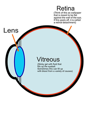
To understand a retinal detachment, think of your eye as a ball with 2 layers - the “shell” which provides the eye with its rigid shape, and the inner layer which is like a thin piece of wallpaper or carpet plastered onto the the shell.
In a retinal detachment, the retina or the wallpaper/carpet comes off the back of the shell causing blurry vision. If left untreated, retinal detachment can lead to permanent vision loss or even blindness.
How a retinal detachment happens


Normal eye (stage 1)
Retinal tear (stage 2)
Retinal detachment (stage 3)
What are the symptoms of a retinal detachment?
Some common symptoms include (please be aware that you may not necessarily experience all of these. There are even some patients who may not notice anything at first!)
-
Sudden increase in floaters: These are small, dark spots or squiggly lines that drift across your vision. While some floaters are normal, a sudden increase can be a sign of retinal detachment.
-
Flashes of light: You may see flashes of light, especially in the periphery of your vision. This can be caused by tears in the retina.
-
Blurred vision: You may experience blurry vision, especially in the area where the retina has detached.
-
Loss of peripheral vision: You may start to lose your vision around the edges of your field of view, often described as a "curtain" or "shadow" coming down over your vision.
-
Sudden vision loss: In some cases, retinal detachment can cause complete vision loss in the affected eye.
What causes a retinal detachment?
The most common cause is aging or as I would say “getting wiser”. As you age, the vitreous within the eye (a sticky gel-like substance) pulls on the retina and causes it to peel off.
Other conditions that increase the risk of a retinal detachment include previous trauma to your head or eye, being short-sighted, or having previous eye surgery such as cataract surgery.
How do you treat a retinal detachment?
The aim of treatment is to reattach the retina to the “shell” of the eye (i.e. fixing the wallpaper back on).
There are 2 main ways it can be done, depending on the type of retinal detachment.
-
Vitrectomy – this is the most common procedure, where we go into the eye, remove any gel (vitreous) that is pulling on the retina, push the retina back against the shell of the eye with a gas or oil bubble, and seal up any holes in the retina with laser.
-
Scleral Buckle – this is a less common procedure but nonetheless has its place for certain types of detachments. For this technique, a “belt” is placed around the eyeball to push the shell of the eye against the retina. In essence, it is about pushing the wall towards the wallpaper.
How successful is treatment?
The chance of reattaching the retina with just one operation is over 90%. This figure does vary depending on the severity of the detachment. Again depending on the detachment, some patients regain full vision, whilst others may notice distortion or deterioration compared to before the detachment. There are lots of nuances and reasons for this, and I would be happy to discuss this in more detail with my patients and their families.
Photo of a retinal detachment
- Blue arrow points to the rip in the retina (retinal tear)
- Orange dotted line demarcates the area of detachment. You can make out that the retina looks raised/ /lifted up/distended in comparison to the rest of the normal retina

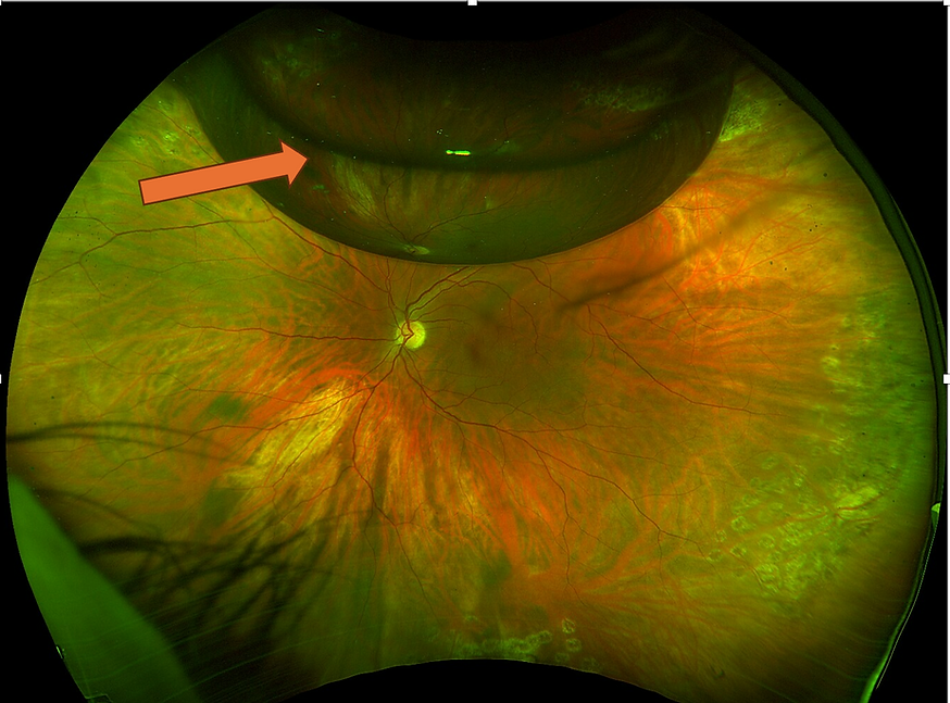
Photo of successful retinal detachment repair with gas
- you can see that the area of detached retina is now flat and looks similar to the rest of the eye.
- the orange arrow points to the gas bubble that I put in your eye. This is approximately 3 weeks after surgery, so the bubble is a lot smaller and doesn't cover the whole eye. It will gradually disappear over time.
What are the risks of surgery?
Any surgery runs the risk of complications such as bleeding, infection and vision loss. If you haven’t had cataract surgery, you will develop a cataract much faster. Cataracts however, are a relatively “easy problem” and can be operated on with good results in almost all cases.
I always tell my patients that there is nothing to lose from surgery for a retinal detachment because the risk of losing your vision without an operation is 100%, compared to a success rate of over 90% if we operate.
Click here for a more detailed discussion on risks of surgery.
What can I expect if I have surgery?
Surgery is usually done under local anaesthetic with some mild sedation i.e. anaesthesia is used to numb the eye whilst some relaxing medication is given via a drip.
All you have to do is lie still on your back, listen to the relaxing music being played whilst the surgery is performed. This takes approximately 45 minutes.
After surgery, a pad is used to cover your eye, and you are then discharged home. We will also give you instructions on how to position after surgery, which may require you to be face-down for a day or 2, followed by sleeping on the left or right side.
What is the aftercare AFTER surgery? When can I go back to work?
I usually see my patients the next day after surgery, and commence eye drops (an antibiotic, a steroid and a dilating drop). You will be reminded to keep the eye clean at all times to avoid the risk of getting an infection. The next appointment will typically be a week after.
I usually advise patients to take at least 1-2 weeks off work and will provide a medical certificate for this. During these 2 weeks, take it easy, avoid any straining or heavy lifting, keep the eye clean, read a book or two, watch some Netflix and go out for nice leisurely walks.
Can I fly after surgery?
It depends. The methods of repairing the retina include either filling the eye with
1) a gas bubble OR
2) an oil bubble
This depends on the type of detachment you have, how long it has been going on for and how "severe" it is. I will discuss this with you after examining you carefully.
If a gas bubble is put in, unfortunately you will not be able to fly until the gas bubble disappears or becomes small enough. This is because gas expands in the eye and cause very high pressures!
On the other hand, if you have an oil bubble in the eye, you will be able to fly. The caveat of having oil in the eye however, is that you will need a second operation to remove the bubble in approximately 2-3 months time.
Can I scuba dive after surgery?
Not immediately! I would avoid any kind of water activities for 2 months to reduce the risk of infections. Once you recover fully and the retina remains stable, (which is about after 2-3 months), you will be able to go back diving if you so wish.